-
Reading Roadmap
- MRI-Based Longitudinal Assessment Predicts Progression to Stage 3 Type 1 Diabetes
- Key Takeaways
- Introduction: The Potential of MRI in Predicting Type 1 Diabetes Progression
- MRI-Based Longitudinal Assessment: A Non-Invasive Approach
- Correlation Between MRI Findings and Clinical Outcomes
- Implications for Treatment and Management
- FAQ Section
- What is type 1 diabetes?
- What are the stages of type 1 diabetes?
- How can MRI scans predict the progression to stage 3 diabetes?
- What are the benefits of early detection?
- Are there any limitations to using MRI for this purpose?
- Conclusion: The Future of Diabetes Management
- Further Analysis
MRI-Based Longitudinal Assessment Predicts Progression to Stage 3 Type 1 Diabetes

[youtubomatic_search]
Key Takeaways
- MRI-based longitudinal assessment can predict the progression to stage 3 type 1 diabetes.
- Early detection of type 1 diabetes progression can lead to better management and treatment strategies.
- Longitudinal MRI scans provide a non-invasive method to monitor pancreatic inflammation and beta-cell loss.
- Research indicates a strong correlation between MRI findings and clinical outcomes in type 1 diabetes patients.
- Further studies are needed to validate these findings and develop standardized protocols for clinical use.
Introduction: The Potential of MRI in Predicting Type 1 Diabetes Progression
Diabetes, specifically type 1 diabetes, is a chronic condition that affects millions of people worldwide. The disease is characterized by the body’s inability to produce insulin due to the autoimmune destruction of beta cells in the pancreas. The progression of type 1 diabetes is typically divided into three stages, with stage 3 being the symptomatic phase requiring insulin therapy. Early detection of progression to stage 3 is crucial for timely intervention and management. Recent research suggests that MRI-based longitudinal assessment could be a promising tool for predicting this progression.
MRI-Based Longitudinal Assessment: A Non-Invasive Approach
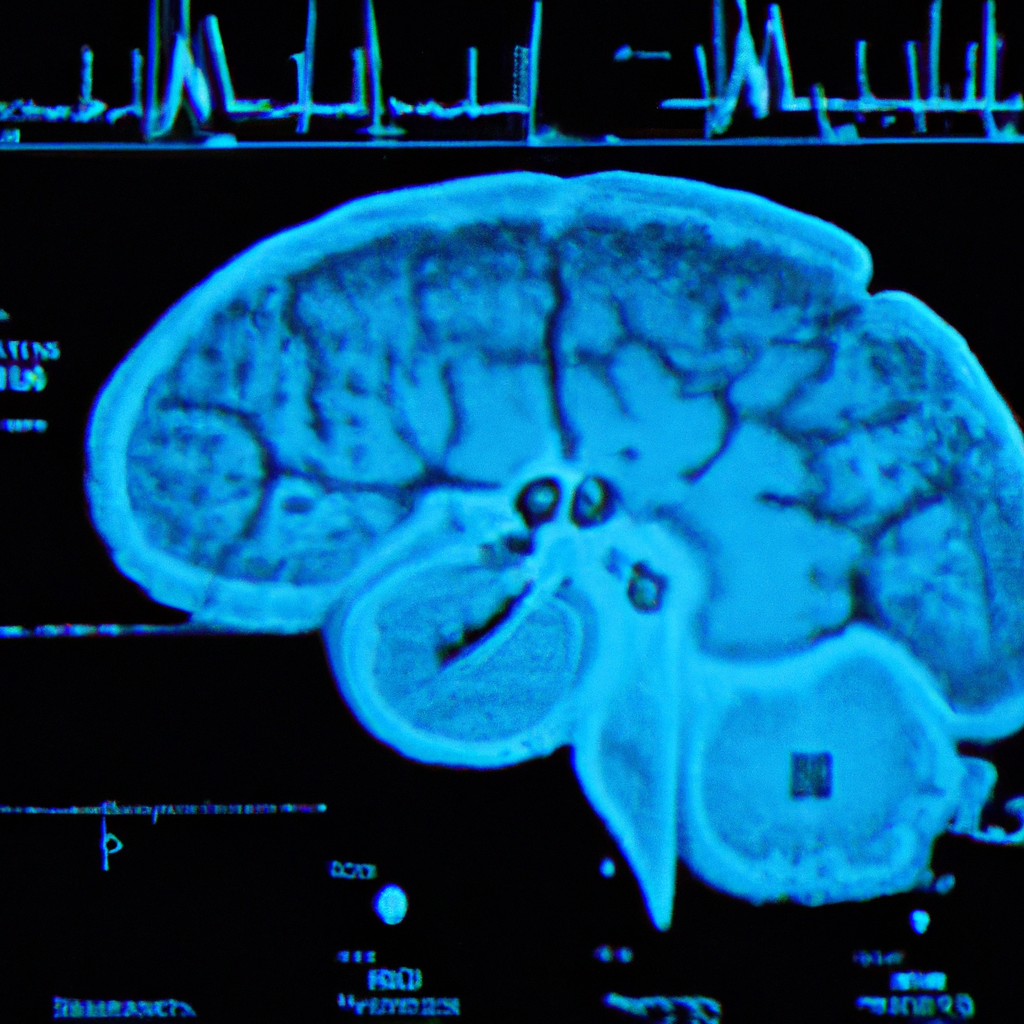
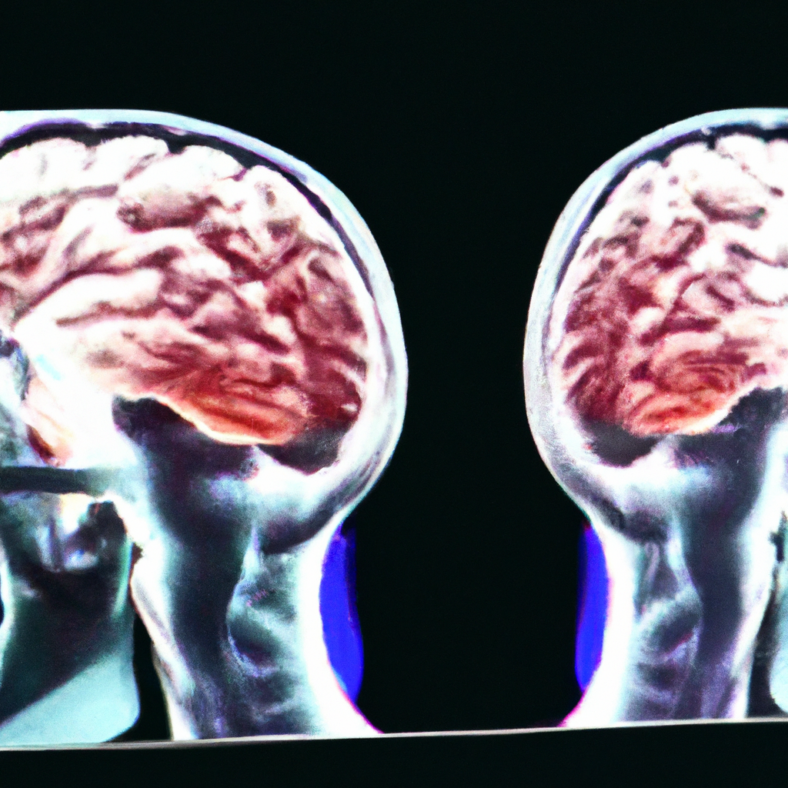
Magnetic Resonance Imaging (MRI) is a non-invasive imaging technique that uses magnetic fields and radio waves to create detailed images of the body’s internal structures. In the context of type 1 diabetes, MRI scans can be used to monitor pancreatic inflammation and beta-cell loss, two key indicators of disease progression. Longitudinal MRI scans, taken over a period of time, can provide valuable insights into how these indicators change and potentially predict the progression to stage 3 diabetes.
Correlation Between MRI Findings and Clinical Outcomes
Several studies have demonstrated a strong correlation between MRI findings and clinical outcomes in type 1 diabetes patients. For instance, a study published in the Journal of Clinical Endocrinology & Metabolism found that patients with higher levels of pancreatic inflammation, as detected by MRI, were more likely to progress to stage 3 diabetes within a year. This suggests that MRI-based longitudinal assessment could be a reliable predictor of disease progression.
Implications for Treatment and Management
The ability to predict the progression to stage 3 diabetes has significant implications for treatment and management. Early detection can allow for timely intervention, potentially delaying the onset of symptoms and reducing the need for insulin therapy. Moreover, it can help clinicians tailor treatment strategies to individual patients, improving outcomes and quality of life.
FAQ Section
What is type 1 diabetes?
Type 1 diabetes is a chronic condition in which the body’s immune system attacks and destroys the insulin-producing beta cells in the pancreas, leading to high blood sugar levels.
What are the stages of type 1 diabetes?
Type 1 diabetes is typically divided into three stages: stage 1 (autoimmunity with no loss of insulin), stage 2 (autoimmunity with abnormal blood sugar levels), and stage 3 (symptomatic diabetes requiring insulin therapy).
How can MRI scans predict the progression to stage 3 diabetes?
MRI scans can monitor pancreatic inflammation and beta-cell loss, two key indicators of type 1 diabetes progression. Longitudinal MRI scans, taken over a period of time, can show how these indicators change and potentially predict the progression to stage 3 diabetes.
What are the benefits of early detection?
Early detection of progression to stage 3 diabetes can allow for timely intervention, potentially delaying the onset of symptoms and reducing the need for insulin therapy. It can also help clinicians tailor treatment strategies to individual patients, improving outcomes and quality of life.
Are there any limitations to using MRI for this purpose?
While MRI-based longitudinal assessment shows promise, further studies are needed to validate these findings and develop standardized protocols for clinical use. Additionally, MRI scans are expensive and may not be accessible to all patients.
Conclusion: The Future of Diabetes Management
The use of MRI-based longitudinal assessment to predict the progression to stage 3 type 1 diabetes represents a significant advancement in the field of diabetes management. This non-invasive approach could provide clinicians with a valuable tool for early detection and intervention, potentially improving patient outcomes and quality of life. However, further research is needed to validate these findings and address the challenges associated with cost and accessibility. As we continue to explore the potential of this technology, we move closer to a future where diabetes management is more personalized, proactive, and effective.
[youtubomatic_search]
Further Analysis
- MRI-based longitudinal assessment can predict the progression to stage 3 type 1 diabetes.
- Early detection of type 1 diabetes progression can lead to better management and treatment strategies.
- Longitudinal MRI scans provide a non-invasive method to monitor pancreatic inflammation and beta-cell loss.
- Research indicates a strong correlation between MRI findings and clinical outcomes in type 1 diabetes patients.
- Further studies are needed to validate these findings and develop standardized protocols for clinical use.

Leave a Reply